Home
Uncategories
Foot Muscles Mri / Mri Of The Ankle Detailed Anatomy W Radiology - This means that the little toe can only be extended by the extensor digitorum longus muscle only.
Foot Muscles Mri / Mri Of The Ankle Detailed Anatomy W Radiology - This means that the little toe can only be extended by the extensor digitorum longus muscle only.
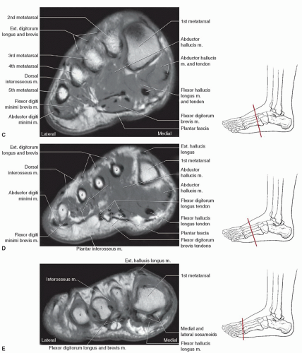
Foot Muscles Mri / Mri Of The Ankle Detailed Anatomy W Radiology - This means that the little toe can only be extended by the extensor digitorum longus muscle only.. 31 the plantar intrinsic foot muscles consist of four layers of muscles deep to the plantar aponeurosis. The flexor digiti minimi brevis (flexor brevis minimi digiti, flexor digiti quinti brevis) lies under the metatarsal bone on the little toe, and resembles one of the interossei. Interestingly the dorsal foot muscles generally have no insertion at the little toe. Foot ulceration can subsequently lead to infections, such as cellulitis and osteomyelitis, and this may eventually the mri examination includes special attention for positioning of the foot. Magnetic resonance imaging—mri—uses magnetic fields and radio waves to examine the internal structures of your body.
It has been widely used to visualise intrinsic muscles because it has high spatial resolution 7, 20, 21, 55, 57, 60. Involved early gray = muscle: There can't be any metal in the room, not just where you have the mri. The muscle that removes the big toe (m.abductor hallucis) lies superficially along the medial edge of the foot. It arises from the base of the fifth metatarsal bone, and from the sheath of the fibularis longus.
Foot Ankle And Calf Musculoskeletal Key from musculoskeletalkey.com What muscle(s) does it originate from and what bone does it insert to? Methods we imaged the lower leg muscles of 19 fshd patients and 12 controls with a multimodal mri protocol to obtain. Mri with hardware in foot? It must be placed in the center of the magnet, to obtain homogeneous fat. Flexion of 4 lesser toes at metatarsophalangeal, proximal & distal interphalangeal joints inversion of foot plantar flexion of ankle. Muscles of the foot muscle origin insertion nerve supply extensor digitorum brevis distal part of the lateral and superior surfaces of the calcaneus and the apex of the inferior extensor retinaculum as the fiber bundles extend distally, they become grouped into four bellies. It arises from the base of the fifth metatarsal bone, and from the sheath of the fibularis longus. Identified 18ffdg pet and mri abnormalities of foot muscles, neurovascular bundles, and skin from 7 crps patient participants.
Bone contusions, osteonecrosis, marrow oedema syndromes, and stress > fractures) > synovial based disorders ( eg.
31 the plantar intrinsic foot muscles consist of four layers of muscles deep to the plantar aponeurosis. Musculoskeletal system | muscle structure and function. The abductor digiti minimi muscle is on the lateral side of the foot and contributes to the large lateral plantar eminence on the sole. Muscle strength) for the foot dorsal and plantar flexors 23. ► shoulder ► elbow ► wrist ► finger ► thumb. This is a 30 year old with swelling on the lateral aspect of foot with evidence of soft tissue lesion in relation to the lateral aspect of the talus which appears isointense to the muscles on t1 and t2 weighted images & appears elongated extending from the anterosuperior calcaneum to the base of. Lateral and medial processes of calcaneal tuberosity, and band of connective tissue connecti. Muscles of the foot are located on its rear and on the sole. Posted by radiologyer at 8:12 am. Human anatomy for muscle, reproductive, and skeleton. Muscles of the ankle and foot. It must be placed in the center of the magnet, to obtain homogeneous fat. There are 10 intrinsic muscles located in the sole of the foot.
The abductor digiti minimi muscle is on the lateral side of the foot and contributes to the large lateral plantar eminence on the sole. Musculoskeletal system | muscle structure and function. It has been widely used to visualise intrinsic muscles because it has high spatial resolution 7, 20, 21, 55, 57, 60. This article reviews the use of magnetic resonance imaging (mri) in the evaluation of the foot, including a discussion of bone and cartilage abnormalities in an article published in the august 2006 issue of this journal, the authors reviewed magnetic resonance imaging (mri) of the ankle. Although almost everyone has muscle cramps at some time, certain factors increase the risk and severity of cramps.
Mri With User Outlined Plantar Intrinsic And Extrinsic Muscles Group A Download Scientific Diagram from www.researchgate.net Where you get the potential for problems with. It has been widely used to visualise intrinsic muscles because it has high spatial resolution 7, 20, 21, 55, 57, 60. Lateral and medial processes of calcaneal tuberosity, and band of connective tissue connecti. Human anatomy for muscle, reproductive, and skeleton. This is a 30 year old with swelling on the lateral aspect of foot with evidence of soft tissue lesion in relation to the lateral aspect of the talus which appears isointense to the muscles on t1 and t2 weighted images & appears elongated extending from the anterosuperior calcaneum to the base of. Although almost everyone has muscle cramps at some time, certain factors increase the risk and severity of cramps. It begins with short tendon bundles on the medial surface of the calcaneus calcaneus, fleshy bundles on the lower retentive flexor. Interestingly the dorsal foot muscles generally have no insertion at the little toe.
Indications for foot mri scan.
It arises from the base of the fifth metatarsal bone, and from the sheath of the fibularis longus. Muscles of the foot are located on its rear and on the sole. What muscle(s) does it originate from and what bone does it insert to? It begins with short tendon bundles on the medial surface of the calcaneus calcaneus, fleshy bundles on the lower retentive flexor. Routine ankle magnetic resonance imaging (mri) tests involve taking images of the foot and ankle in the axial, coronal, and sagittal planes the imaging process allows the magnetic field to find changes in the organ and tissue structures, identifying any sprains, ruptures, dislocations, or synovial disorders. Start studying mri procedures foot/ankle review. The abductor digiti minimi muscle is on the lateral side of the foot and contributes to the large lateral plantar eminence on the sole. In addition, an image of all the muscles of the back and plantar part of the foot, all tendons and tendon ligaments, blood vessels and nerves are obtained. Magnetic resonance imaging (mri) is the method of choice for detecting soft tissue structure and abnormalities 58, 59. The flexor digiti minimi brevis (flexor brevis minimi digiti, flexor digiti quinti brevis) lies under the metatarsal bone on the little toe, and resembles one of the interossei. ► shoulder ► elbow ► wrist ► finger ► thumb. It must be placed in the center of the magnet, to obtain homogeneous fat. Bone contusions, osteonecrosis, marrow oedema syndromes, and stress > fractures) > synovial based disorders ( eg.
Routine ankle magnetic resonance imaging (mri) tests involve taking images of the foot and ankle in the axial, coronal, and sagittal planes the imaging process allows the magnetic field to find changes in the organ and tissue structures, identifying any sprains, ruptures, dislocations, or synovial disorders. It begins with short tendon bundles on the medial surface of the calcaneus calcaneus, fleshy bundles on the lower retentive flexor. The muscle that removes the big toe (m.abductor hallucis) lies superficially along the medial edge of the foot. They act collectively to stabilise the arches of the foot, and individually to control movement of the digits. Identified 18ffdg pet and mri abnormalities of foot muscles, neurovascular bundles, and skin from 7 crps patient participants.
Denervation Pseudohypertrophy Of Calf Muscles Associated With Diabetic Neuropathy Sciencedirect from ars.els-cdn.com Mr data were then acquired. Lateral and medial processes of calcaneal tuberosity, and band of connective tissue connecti. This article reviews the use of magnetic resonance imaging (mri) in the evaluation of the foot, including a discussion of bone and cartilage abnormalities in an article published in the august 2006 issue of this journal, the authors reviewed magnetic resonance imaging (mri) of the ankle. Where you get the potential for problems with. In addition, an image of all the muscles of the back and plantar part of the foot, all tendons and tendon ligaments, blood vessels and nerves are obtained. The muscles acting on the foot can be divided into two distinct groups; This means that the little toe can only be extended by the extensor digitorum longus muscle only. The purpose of this study was to investigate the relationship of muscle mri findings and gait disturbance in myotonic dystrophy type 1 (dm1) patients.
A magnetic resonance imaging (mri) was performed on a normal subject;
It begins with short tendon bundles on the medial surface of the calcaneus calcaneus, fleshy bundles on the lower retentive flexor. Posted by radiologyer at 8:12 am. Where you get the potential for problems with. Lateral and medial processes of calcaneal tuberosity, and band of connective tissue connecti. This is a 30 year old with swelling on the lateral aspect of foot with evidence of soft tissue lesion in relation to the lateral aspect of the talus which appears isointense to the muscles on t1 and t2 weighted images & appears elongated extending from the anterosuperior calcaneum to the base of. Synovitis, tenosynovitis, bursitis, and ganglion cysts) > congenital and developmental conditions( eg.dysplasia, tarsal coalition). Involved early gray = muscle: Mri of the soft tissues of the foot visualizes the fat cushions of the sole, heels, fingers and can show swelling, foci of infiltration and inflammation. Although almost everyone has muscle cramps at some time, certain factors increase the risk and severity of cramps. Start studying mri procedures foot/ankle review. Human anatomy for muscle, reproductive, and skeleton. Routine ankle magnetic resonance imaging (mri) tests involve taking images of the foot and ankle in the axial, coronal, and sagittal planes the imaging process allows the magnetic field to find changes in the organ and tissue structures, identifying any sprains, ruptures, dislocations, or synovial disorders. Mri with hardware in foot?


0 Comments:
Posting Komentar